

.gif)
Didi u know about SKIN LESIONs (kudhurika kwa Ngozi)
Did u know about Skin lesion?; know then SAY NO TO STIGMATIZATION
 my dear to day am giving you what you are supposed to know about your skin, for a good health of your skin you should know these kind of lesion,this will be easily for you to take medication and to make your life happy:A skin lesion is a zone of tissue with impaired function as a result of damage by disease or wou
nding.(vidonda)
skin lesion cab be Classified as primary or secondary lesion
to day i will start with Primary lesions:
Primary lesions are either the first visible lesion or involve the initial skin changes.
Examples:
1. A macule is a flat, non-palpable lesion, variably shaped,
discoloured skin lesion of less than 10 mm in diameter.
2. A patchy is a large macule of more than 10 mm in diameter.
 Examples:
 4. A papule is a solid, elevated lesion usually of less than 10 mm in diameter.
5. A nodule is a palpable,

solid lesion of 10-20 mm in diameter e.g. molluscum contagiosum and chicken pox lesions.
6. A tumour (swelling) is a larger nodule of more than 20 mm in diameter.
 • Size.
9. Pustules are small, circumscribed skin papules containing purulent material that is less than 5 mm in diameter.


13. Angioneurotic oedema is larger localized areas of oedema in the subcutaneous tissue.

 Purpura is a condition of red or purple discoloured spots on the skin that do not blanch on applying pressure.
 o Petechiae are not found on the sole of the foot where the vessels are protected by the strong subcutaneous tissue.
15. Comedones:
17. Erythema:
This is reddening of the skin due to the dilatation of blood capillaries just below the epidermis in the dermis. In black individuals, this appears as shining skin.

2. Lichenification is a thickened skin area with accentuated skin markings.

 6. Fissure is a deep skin split extending into the dermis.
7. Erosion is a superficial, focal loss of part of the epidermis. Lesions usually heal without scarring.

10. Scar is an abnormal fibrous tissue that replaces normal tissue after skin injury.

A scar can either be:
• Hypopigmentation is a decreased skin pigment as in Tinea versicolor.


• Hyperpigmentation is an increased skin pigment as in haemochromatosis.
Pattern of distribution and their differential diagnosises
 my dear to day am giving you what you are supposed to know about your skin, for a good health of your skin you should know these kind of lesion,this will be easily for you to take medication and to make your life happy:A skin lesion is a zone of tissue with impaired function as a result of damage by disease or wou
nding.(vidonda)
skin lesion cab be Classified as primary or secondary lesion
to day i will start with Primary lesions:
Primary lesions are either the first visible lesion or involve the initial skin changes.
Examples:
1. A macule is a flat, non-palpable lesion, variably shaped,
discoloured skin lesion of less than 10 mm in diameter.
2. A patchy is a large macule of more than 10 mm in diameter.
Examples:• Tattoos.• Freckles.• Flat moles.• Port-wine stains.• Fixed drug reaction.
3. A plaque is a plateau-like lesion of more than 10 mm or a group of confluent papules.
 Examples:
• Warts.
• Psoriasis.
• Insect bites.
• Some naevi.
• Lichen planus.
• Syphilitic chancre.
 4. A papule is a solid, elevated lesion usually of less than 10 mm in diameter.
solid lesion of 10-20 mm in diameter e.g. molluscum contagiosum and chicken pox lesions.
6. A tumour (swelling) is a larger nodule of more than 20 mm in diameter.

• Site.
• Shape.
• Surface.
• Shifting (movements).
• Softness (consistency).
• Smoothness (regularity).
Examples:• Lipoma.• Fibroma.• Keratinous cysts.• Erythema nodosum.
7. Cysts

are enclosed cavities with a lining that can contain a liquid or semisolid material.

are enclosed cavities with a lining that can contain a liquid or semisolid material.

Causes:
• Rosacea.
• Idiopathic.
• Long-term therapy with topical fluorinated corticosteroids.
• Certain systemic diseases e.g. ataxia-telangiectasia or scleroderma.
• Inherited disorders e.g. hereditary ataxia-telangiectasia and hereditary haemorrhagic telangiectasia.
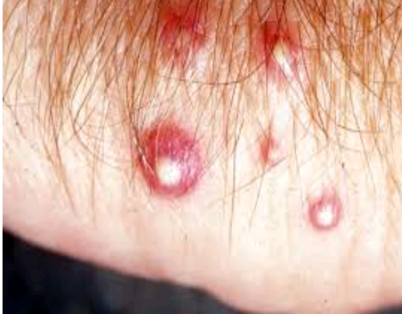
10. A vesicle is a circumscribed, skin papules containing serous fluid that is less than 5 mm in diameter.

11. A bulla (blister) is a vesicle or pustule of more than 5 mm in diameter.


Causes:
• Sunburn.
• Pemphigus.
• Insect bites.
• Pemphigoid.
• Drug eruptions.
• Physical trauma.
• Primary irritants.
• Epidermolysis bullosa.
• Erythema multiforme.
• Dermatitis herpetiformis.
• Allergic contact dermatitis.
• Viral infections e.g. herpes simplex or herpes zoster.
12. Wheals (hives) are transient, elevated lesions caused by localized oedema.


Causes: Wheals are a common allergic reaction e.g.
• Insect bites.
• Drug eruptions.
• Sensitivity to cold, heat, pressure or sunlight.
13. Angioneurotic oedema is larger localized areas of oedema in the subcutaneous tissue.

14. Purpura:
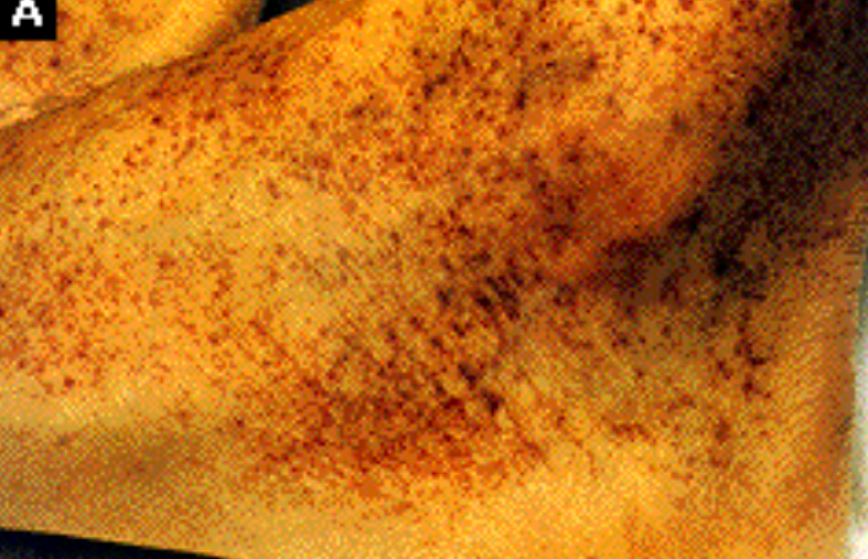 Purpura is a condition of red or purple discoloured spots on the skin that do not blanch on applying pressure.
The spots are caused by bleeding underneath the skin usually secondary to vasculitis or dietary deficiency of vitamin C (scurvy).
• Petechiae:
o Petechiae are small (1–2 mm) red or purple spot on the skin, caused by a minor bleed from broken capillary blood vessels.
o They characteristically develop in crops in areas of increased venous pressure, such as the dependent parts of the body. As a result, they are most dense on the feet and ankles, fewer are present on the legs.
 o Petechiae are not found on the sole of the foot where the vessels are protected by the strong subcutaneous tissue.
• Ecchymosis: It is larger confluent areas of extravasations.
• Haematoma is an area of massive bleeding into the skin and underlying tissues.
15. Comedones:
• Definition:
They are bumps resulting from a collection of sebum, keratinised cells and waste which accumulate in the entrance of the follicle.

• Types:
o Open comedones:
Open comedones ‘blackhead’ bumps contained within the follicle.
o Closed comedones:
Closed comedones are white headed-bumps, trapped underneath the skin’s surface.
16. Crow’s feet:
• Definition:
These are fine lines around the eyes caused by habitual facial expressions and daily movement.


• Cause:
They are associated with ageing of muscle tissues but premature formation may be due to over exposure to ultraviolet light or eye strain.
17. Erythema:
This is reddening of the skin due to the dilatation of blood capillaries just below the epidermis in the dermis. In black individuals, this appears as shining skin.

18. Keloid:


• A keloid is the over growth of an existing scar which grow much larger than the original wound.
• The surface may be smooth, shiny or ridged. The onset is gradual and is due to an accumulation or increase in collagen in the immediate area.
• The colour varies from red, fading to pink and white.
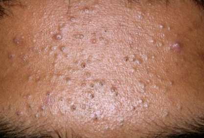
19. Milia:


• Definition: Milia are sebum-rich bumps trapped in a blind duct with no surface opening.
• They are usually around the eye area; they appear as pearly, white and hard nodules under the skin.

• They may have hairs growing on them.
• They appear as round, smooth lumps on the surface of the skin.
• They may be flat or raised, vary in size and colour from pink to brown or black.
21. Skin tag:

They are small growths of fibrous tissue, which stand up from the skin.

They are small growths of fibrous tissue, which stand up from the skin.
Secondary lesions:
Secondary lesion is a lesion that results when primary lesions that undergo a natural evolution.
• Examples:
o A burst vesicle.
o Infected lesion.
o Manipulated lesion e.g. a scratched vesicle.
Examples:
1. Excoriation is a linear or hollowed-out crusted area caused by scratching, rubbing or picking.


2. Lichenification is a thickened skin area with accentuated skin markings.

Causes:
• Onchocerciasis.
• Atopic dermatitis.
• Lichen simplex chronicus (localized scratch dermatitis).

4. Scales are superficial epidermal cells that are dead and cast off from the skin.


Scaling rashes:
• Psoriasis.
• Pityriasis rosea.
• Tinea versicolor.
• Seborrhoeic dermatitis.
• Superficial fungal infections.
• Chronic dermatitis of any type.
5. Crust (scab) is a dried serum, blood or pus.


Causes:
• Infectious diseases.
• Inflammatory diseases.
 6. Fissure is a deep skin split extending into the dermis.
7. Erosion is a superficial, focal loss of part of the epidermis. Lesions usually heal without scarring.

Causes:
• Pemphigus.
• Tinea pedis.
• Herpes viruses.
8. An ulcer is focal loss of the epidermis extending into the dermis.


Causes:
• Tumours.
• Neuropathies.
• Physical trauma.
• Systemic scleroderma.
• Acute bacterial infection.
• Peripheral vascular diseases.
• Chronic bacterial and fungal infection.
9. Atrophy is a decreased skin thickness due to skin thinning.


Causes:
• Ageing.
• Sometimes after burns.
• Discoid lupus erythematosus.
• Long-term use of topical potent corticosteroids.
10. Scar is an abnormal fibrous tissue that replaces normal tissue after skin injury.

A scar can either be:
• Cosmetic scar.
• Traumatic scars.
• Therapeutic scar.
Causes:
• Cuts.
• Burns.
• Diseases such as discoid lupus erythematosus.
11. Pigmentation:


• Depigmentation is total loss of skin pigment like in Vitiligo.
• Hypopigmentation is a decreased skin pigment as in Tinea versicolor.


• Hyperpigmentation is an increased skin pigment as in haemochromatosis.
Pattern of distribution and their differential diagnosises
1. Flexural area• Atopic dermatitis.• Bullous pemphigoid.• Acanthosis nigricans.
2. Extensor surface • Psoriasis.• Xanthomas.• Atopic dermatitis.• Dermatitis .
3. Feet and hands • Scabies.• Eczema.• ID reaction.• Tinea infection.• Erythema
4. Wrist and ankle • Scabies.• Eczema. • Lichen planus.• Contact dermatitis.
5. Sun-exposed skin areas • Pellagra.• Dermatomyositis.• Photo-drug eruption.• Porphyria cutanea tarda.• Polymorphous light eruption.• Systemic Lupus
6. Mouth • Leukoplakia.• Skin cancers.• Mucous cysts.• Fordyce spots.• Kaposi’s s
7. Axillae • Impetigo.• Folliculitis.• Erythrasma.• Achrocordon.• Contact dermatitis.• Acanthosis nigricans.• Hailey-Hailey disease.•
8. Buttocks • Scabies.• Psoriasis.• Folliculitis.• Kawasaki disease.• Streptococcal cellulitis.• Hidradenitis suppurativa.• Lichen sclerosus et atrophicus.
9. Scalp • Kerion.• Psoriasis.• Tinea capitis.• Discoid lupus.• Contact dermatitis.• Seborrhoea dermatitis.
No comments
Post a Comment